Publications
Sharing our work!
Select Publications
-

MRI in the Evaluation of Cryptogenic Stroke and Embolic Stroke of Undetermined Source (Apr 2024)
We review the diagnostic utility of various MRI techniques in patients with cryptogenic stroke.
-

Vessel wall imaging in the diagnosis of antiphospholipid syndrome presenting as Moyamoya syndrome (Apr 2024)
We report a fascinating case of APLS vasculopathy with vessel wall MR imaging findings.
-

Implementation of a Clinical Vessel Wall MR Imaging Program at an Academic Medical Center (Mar 2024)
We describe how to use principles of implementation science and the RE-AIM framework to succssfully build a clinical vessel wall imaging program at an academic medical center.
-

Ophthalmic Artery Vessel Wall Inflammation in a Patient With Giant Cell Arteritis Presenting With Vision Loss (Mar 2024)
We report scalp and orbital MR findings in a patient with giant cell arteritis with inflammation of the temporal and ophthalmic arteries.
-

Sex Differences in Perihematomal Edema Volume and Outcome After Intracerebral Hemorrhage (Feb 2024)
In this post hoc analysis of the Factor VIIa for Acute Hemorrhagic Stroke Treatment (FAST) trial, early PHE expansion and trajectory in men were significantly higher than women.
-

Emerging Concept of Intracranial Arterial Diseases: The Role of High Resolution Vessel Wall MRI (Jan 2024)
We review the embryology of the intracranial arteries, spectrum and vessel wall imaging appearances of intracranial arterial diseases and highlight future research directions on understanding the temporal profile of VWI findings and developing quantitative interpretative approaches.
-
High-resolution Magnetic Resonance Imaging Visualizes Intracranial Large Artery Involvement in Giant Cell Arteritis (Jan 2024)
In this multicenter study evaluating patients with GCA and imaged with VWI, up to 14.5% of patients showed intracranial inflammatory vessel wall changes.
-

Imaging Biomarkers & Prevalence of Complex Aortic Plaque in Cryptogenic Stroke (Nov 2023)
We review imaging features by imaging modality (TEE, CT/CTA, MR/MRA) of complex aortic plaque. The prevalence of complex aortic plaque is also calculated in patients with embolic stroke of undetermined source. See twitter teaser here.
-

Carotid Plaque-RADS: A Novel Stroke Risk Classification System (Oct 2023)
In this international collaborative work, we report a new Plaque-RADS nomenclature to classify carotid plaque types based on composition and morphology via different imaging modalities, such as US, CT, and MRI.
-

Streamlined Carotid Artery Calcification Labeling For CTA Scans (July 2023)
We report the speed and accuracy of a novel web-based software for streamlined single-click calcific plaque labeling compared to manual segmentation using 3D slicer. (Open Source)
-
MR Imaging for Intracranial Vessel Wall Imaging: Pearls and Pitfalls (May 2023)
We review the clinical applications for intracranial VWI and interpretive pearls and technical pitfalls for this innovative imaging technique.
-
Orbital magnetic resonance imaging of giant cell arteritis with ocular manifestations: a systematic review and individual participant data meta-analysis (May 2023)
We systematically review all orbital MRI findings of patients with biopsy-proven GCA and visual disturbances. We report the clinical/exam findings and prevalence of orbital imaging findings in this disease.
-

Diagnostic Modalities in Giant Cell Arteritis (Apr 2023)
We review diagnostic tests used for giant cell arteritis, including imaging, lab, and histopathology.
-

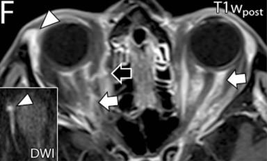
Intraorbital Findings in Giant Cell Arteritis on Black Blood MRI (Nov 2022)
This multicenter study examines orbital MRI findings using black blood (vessel wall) MRI in patients with Giant Cell Arteritis. Figure shows enhancement of the ophthalmic artery using VW-MRI (arrows).
-
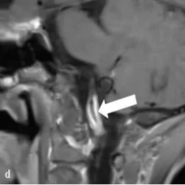
Vessel Wall Magnetic Resonance Imaging of a Nonstenotic Craniocervical Vertebral Artery Dissection (Sept 2022)
We report how VWI can facilitate diagnosis of a craniocervical vertebral artery dissection.
-

Vessel Wall MR Imaging of Aortic Arch, Cervical Carotid and Intracranial Arteries in Patients with Embolic Stroke of Undetermined Source (July 2022)
We review intracranial, carotid, and aortic vessel wall MR imaging techniques used to identify culprit plaque in patients with ESUS. View animated visual abstract here.
-

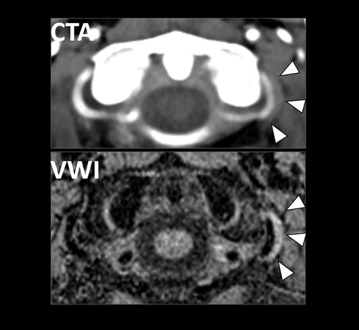
Vessel Wall Imaging of a Carotid Web (May 2022)
We report imaging findings on CTA and VWI of a carotid web that was the most likely cause of ischemic stroke.
-

Sex Differences in Intracranial Atherosclerosis in Patients With Hypertension With Acute Ischemic Stroke (May 2022)
This multicenter (China and USA) retrospective study evaluates sex-differences in intracranial plaque burden in hypertensive patients with acute ischemic stroke.
-

Current Clinical Applications of Intracranial Vessel Wall MR Imaging (Oct 2021)
We review vessel wall MR imaging (VWI) features of different intracranial vasculopathies and show illustrative clinical cases.
-

Cardiovascular Risk Factors Affect Specific Segments of the Intracranial Vasculature in High-Resolution Vessel Wall Imaging (Oct 2021)
In collaboration with Dr. Javier Romero’s team at MGH/HMS, this work used high-resolution vessel wall imaging to determine associations between cardiovascular risk factors and specific intracranial vessel segment involvement.
-
Diagnostic Accuracy Studies in Radiology: How to Recognize and Address Potential Sources of Bias (Sept 2021)
Accuracy is an important parameter of a diagnostic test. We provide a framework for evaluating potential sources of study design biases that are found in diagnostic accuracy studies and explain their impact on sensitivity and specificity estimates.
-

Imaging endpoints of intracranial atherosclerosis using vessel wall MR imaging: a systematic review (Jun 2021)
We systematically review the vessel wall MR imaging literature to assess the criteria and measurement methods of VWI-related imaging endpoints for symptomatic intracranial plaque in patients with ischemic events.
-

Sex Differences in Carotid Plaque Composition in Patients With Embolic Stroke of Undetermined Source (May 2021)
We examined sex differences in nonstenotic carotid plaque composition in patients with embolic stroke of undetermined source.
-

Vessel wall MR imaging of central nervous system vasculitis: a systematic review (May 2021)
We systematically reviewed the literature on vessel wall MR imaging patterns of inflammatory versus infectious vasculitis and also compared imaging patterns for intracranial versus extracranial arteries of the head and neck.
-

Detection of Angiographically Occult Ruptured Basilar Sidewall Perforator Aneurysm by Vessel Wall MR Imaging (Apr 2021)
We report a case that highlights the role vessel wall MR imaging can play in detecting small ruptured aneurysms that intermittently thrombose and are otherwise challenging to diagnose with conventional vessel imaging.
-

Vessel Wall Imaging of Basilar Artery Perforator Disease (Mar 2021)
We report a case that highlights the utility of vessel wall MR imaging in detecting nonstenotic basilar artery plaque contributing to an acute pontine infarct.
-

Vessel Wall Magnetic Resonance Imaging Biomarkers of Symptomatic Intracranial Atherosclerosis: A Meta-Analysis (Jan 2021)
We performed a meta-analysis to evaluate the strength of association of imaging features of symptomatic plaque leading to downstream ischemia.
-

Intracranial vessel wall MR imaging of an intradural vertebral artery dissection (Dec 2020)
We report a case highlighting the role of high-resolution vessel wall MR imaging as a useful adjunct to conventional lumen-based imaging techniques for diagnosing arterial dissections.
-

Emerging multi-modal diagnostic approaches for moyamoya disease (Oct 2020)
We review Ya et al’s work entitled, “High-resolution combined arterial spin-labeling MR for identifying cerebral arterial stenosis induced by moyamoya disease or atherosclerosis.”
-

Data-Driven Quantitative Susceptibility Mapping Using Loss Adaptive Dipole Inversion (LADI) (Sept 2020)
For neuroimaging applications, loss adaptive dipole inversion (LADI) preserved image sharpness and fine features in patients with glioblastoma and microbleeds. LADI performed better at mitigating artifacts in cardiac QSM. This work was in collaboration with and led by members of Dr. Walter Witschey’s CorLab.
-

Vessel wall MR imaging of intracranial atherosclerosis (Aug 2020)
In this review, we discuss the advantages of intracranial vessel wall MR imaging for intracranial atherosclerosis, the potential of bioimaging markers to identify vulnerable intracranial plaque, and future directions of artificial intelligence and its utility for lesion scoring and assessment.
-

MR Intracranial Vessel Wall Imaging: A Systematic Review (July 2020)
We systematically review the literature to identify trends and extent of variability in intracranial vessel wall MR imaging techniques and protocols.
-

Qualitative Assessment and Reporting Quality of Intracranial Vessel Wall MR Imaging Studies: A Systematic Review (Dec 2019)
We performed a systematic review of publication trends and reporting quality of VWI publications to identify unmet needs and future directions.
-

Spatial Distribution of Intracranial Vessel Wall Enhancement in Hypertension and Primary Angiitis of the CNS (Dec 2019)
Sspatial distribution of vessel wall enhancement is an important consideration when interpreting vessel wall MR exams, particularly in patients with a comorbid diagnosis of hypertension.
-

Serial vessel wall MR imaging of pediatric tuberculous vasculitis (Dec 2019)
This case illustrates the radiologic evolution of tuberculous vasculitis by serial vessel wall imaging,
-

Central Nervous System Vasculopathies (Nov 2019)
We review the salient clinical and imaging features of noninflammatory and inflammatory CNS vasculopathies.
-

Vessel Wall MRI Added to MR Angiography in the Evaluation of Suspected Vasculopathies (July 2019)
3D-TOF-MRA and vessel wall MR imaging together identifies a higher number of abnormal vessel segments than 3D-TOF-MRA alone and may provide a more accurate assessment of disease burden.
-

High Resolution Vessel Wall MR imaging in prestenotic intracranial atherosclerotic disease (May 2019)
We present a case of intracranial hemorrhage presumably due to intracranial atherosclerotic disease identified by vessel wall MR imaging and silent on lumen-based imaging modalities.
-

High Resolution Vessel Wall MRI and Vasculopathy Related to Herpes Zoster Ophthalmicus (Jul 2018)
We show a case of a patient with VZV vasculitis imaged with high resolution vessel wall magnetic resonance imaging. There was circumferential wall thickening and enhancement of the infected vessels.